Isotype Controls: An Important Experimental Tool

Table of Contents
- Overview
- What Are Antibody Isotypes?
- What is an Isotype Control?
- Do I Need to Use Isotype Controls?
- What Causes Background Staining?
- How to Choose an Isotype Control
- Analyzing Isotype Control Results
- Use of Isotype Controls in Vivo
- Isotype Controls for Human Studies
- References
Overview
Anti-body-based experimental design requires the use of appropriate controls. The use of inadequate controls confound data interpretation and reduce the likelihood of obtaining meaningful results. Antibody isotype controls are essential negative controls for in vitro and in vivo studies and are used extensively in applications such as flow cytometry, immunohistochemistry, immunocytochemistry, enzyme-linked immunosorbent assays (ELISAs) and western blotting. This article explains the purpose for isotype controls and why they are an essential part of antibody-based experiments.
What are Antibody Isotypes?
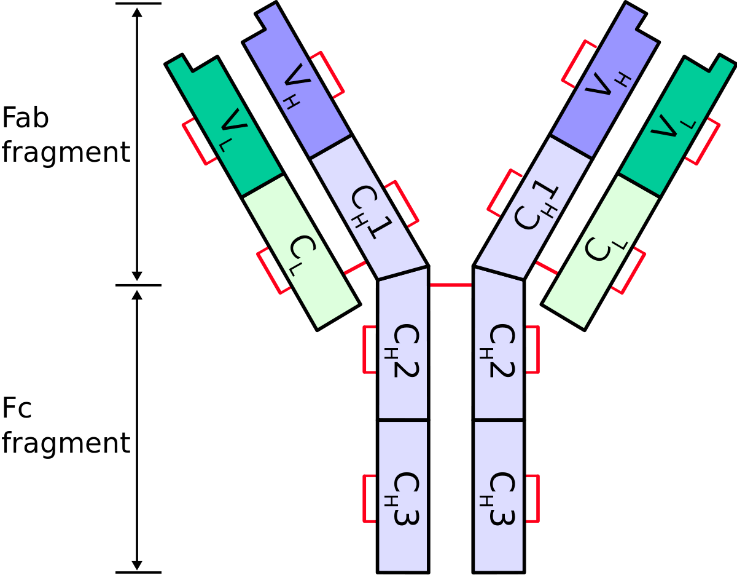
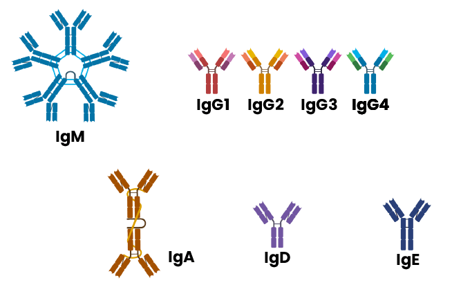
To understand what an antibody isotype is, we first need to explore the main structure of an antibody (also known as an immunoglobulin). Antibodies are Y-shaped proteins composed of two identical light chains and two identical heavy chains linked by disulfide bonds (Figure 1a). There are two types of light chain: κ and λ, and five types of heavy chain: α, γ, δ, ε, and μ. This latter group of heavy chains serves as the basis for distinguishing five antibody classes or isotypes: IgA, IgG, IgD, IgE, and IgM, respectively. IgA and IgG may be further subdivided based on heavy chain differences into IgA1 or IgA2, and IgG1, IgG2, IgG3 and IgG4. Light chains consist of a constant domain and a variable domain, while heavy chains consist of either three constant domains and one variable domain (IgA, IgD, and IgG) or four constant domains and one variable domain (IgE and IgM). Most isotypes are monomeric and thus consist of a single pair of heavy-light chains (IgG, IgD, IgE), while IgA and IgM have a structure enabling dimeric and pentameric heavy-light chain configurations (Figure 1b). [1, 2]


Figure 1. Typical antibody structure and different antibody isotypes.
The functional composition of an antibody consists of two fragment antigen binding domains (Fab) and a fragment crystallisable domain (Fc) (Figure 1a). The variable domains of the Fab (Fv) form the antigen binding site. The Fab fragment is bound via a hinge region to the Fc, which in turn is responsible for binding to receptors and determining the effector function profile of the antibody.[1] These functions include recruitment of immune cells that recognise the isotype Fc, binding to complement system proteins, and transportation of antibodies through cells and into different tissues. [3]
Isotypes differ in their biological function, physiological location, and ability deal with different antigens during the phases of an immunological response.[2-4] IgM is produced during the primary immune response (initial interact with antigens to activate B cells), while IgG is the most produced isotype during the secondary immune response (when antigens are re-encountered). IgE has a role in anaphylaxis and parasite protection. IgA is the most prevalent isotype in body secretions (e.g. saliva, milk, intestinal and bronchial secretions) where it helps to protect mucosal membranes. The function of IgD remains unknown.[2-4] Further detail on the function and distribution of each isotype is presented in Table 1.
| IgM | IgG1 | IgA | IgE | IgD | |
| Function | |||||
| Neutralization | |||||
| Opsonization | |||||
| Sensitization for Killing by NK Cells | |||||
| Sensitization of Mast Cells | |||||
| Activates Complement System | |||||
| Distribution | |||||
| Transport Across Epithelium | |||||
| Transport Across Placenta | |||||
| Diffusion into Extravascular Sites |
Table 1. Function and distribution profile for antibody isotypes. [4]
Red – no function/distribution role.
Dark green – major effector/distributor function.
Mid-green – lesser function/distribution.
Light green – very minor function/distribution.
What is an Isotype Control?
When conducting antibody-based experiments, it is useful to know the antibody isotype in order to select the matching isotype control. An isotype control has the same constant heavy chain as the primary antibody isotype but lacks the ability to bind the target antigen. This implies that an isotype control may be used as a negative control to help demonstrate that the reaction of interest is indeed due to the genuine interaction of the antigen epitope and the primary antibody. [5] In turn, isotype controls allow us to determine the contribution of non-specific background activity and normalise for it when measuring the true signal of the interaction under study.
Do I Need to Use Isotype Controls?
Background staining (an indicator of non-specific reactivity between the primary antibody and Fc receptors on various cells) is common in applications such as flow cytometry and immunohistochemistry. The use of isotype controls in these applications, and other experiments where background reactivity may interfere with the results, is imperative for rigorous experimental design and reliable data interpretation. Isotype controls serve as essential negative controls to account for non-specific background staining signals and thus distinguish specific from non-specific antibody binding. The inclusion of isotype controls strengthens the specificity of results by enabling discernment of true positive signal.
What Causes Background Staining?
Although tissue-dependent, background staining can be attributed to non-specific antibody binding to Fc receptors across a variety of cells. [6] Strong background staining may also be caused by interference from endogenous peroxidases or phosphatases, unblocked endogenous biotin or lectins, and nonspecific interactions between the primary antibody and non-target epitopes. [7]
How to Choose an Isotype Control
Selecting an optimal isotype control involves matching key primary antibody characteristics such as host species, isotype (and subclass if appropriate), and type of conjugation. If, for example, a Mouse IgG1 is used as the primary antibody then a Mouse IgG1 isotype control should be used as the isotype control. Ichorbio offers a variety of isotypes controls to match your experimental needs; all products are of high purity and have low endotoxin levels, low protein aggregation, and are pathogen free (visit our products page here).
Analyzing Isotype Control Results
Compare signal from primary antibody to isotype control run under same conditions. Minimal staining indicates low background. Considerable isotype control signal reveals background level to interpret actual antibody binding signal.
While isotype controls reveal background staining, they don't confirm antibody specificity or indicate background source. Still, they are an essential control for reliable immunology experiments.
Additionally, utilize the same experimental conditions between the paired antibodies like concentration, incubation parameters, blocking solutions, and detection methods for valid comparisons. Matching these critical properties while minimizing procedural differences allows the isotype control to act as an accurate negative surrogate for interpreting specific binding. With careful antibody characterization and controlled testing conditions, the isotype control reliably models background noise, enabling specificity determinations.
In summary, isotype controls are an essential experimental tool for immunology studies. They help control for non-specific background signal and antibody effects like HAMA interference. Careful selection of the appropriate isotype control for each target antibody and sample type allows you to confidently interpret the specific binding in your experiments. Isotype controls provide an additional level of scientific rigor that produces reliable immunology data.
Uses of Isotype Controls in Vivo
When performing in vivo experiments, isotype controls are extremely useful for analyzing cells and tissues by flow cytometry or microscopy. Tissues contain many proteins that antibodies can bind to non-specifically. Using an isotype control antibody helps determine the level of background signal in your experiment. Samples labeled with the isotype control can then be compared to those labeled with the specific antibody. Any signal greater than the isotype control is likely due to specific binding of the target antibody.
Isotype Controls for Human Studies
For experiments involving human samples, choosing the right isotype control is critical. Since the test antibody and isotype control are both mouse antibodies, they could cause false positive signals by binding to human anti-mouse antibodies (HAMA) present in patient samples. Using a mouse antibody with the same isotype but different specificity helps control for these effects. An IgG1 control is standard for human IgG1 antibodies, but for other subtypes like IgG2 or IgM, an antigen-specificity matched control provides even better specificity. With the right isotype-matched controls, you can effectively identify specific target antibody binding in human samples.
References
- Chiu, M.L., et al., Antibody Structure and Function: The Basis for Engineering Therapeutics. Antibodies (Basel), 2019. 8(4).
- Cruse, J.M. and R.E. Lewis, Atlas of Immunology. Third ed. 2010, Boca Raton, FL.: CRC Press. Taylor & Francis Group.
- Kenkel, B. Antibodies 101: Isotypes. 2021 February 2024]; Available from: https://blog.addgene.org/antibodies-101-isotypes
- Janeway, C.A.J., et al., The distribution and functions of immunoglobulin isotypes. 5th ed. Immunobiology: The Immune System in Health and Disease. 2001, New York: Garland Science.
- Hewitt, S.M., et al., Controls for immunohistochemistry: the Histochemical Society's standards of practice for validation of immunohistochemical assays. J Histochem Cytochem, 2014. 62(10): p. 693-7.
- Buchwalow, I., et al., Non-specific binding of antibodies in immunohistochemistry: fallacies and facts. Sci Rep, 2011. 1: p. 28.
- Scientific., T. IHC Troubleshooting Guide. February 2024]; Available from: https://www.thermofisher.com/uk/en/home/life-scie...


