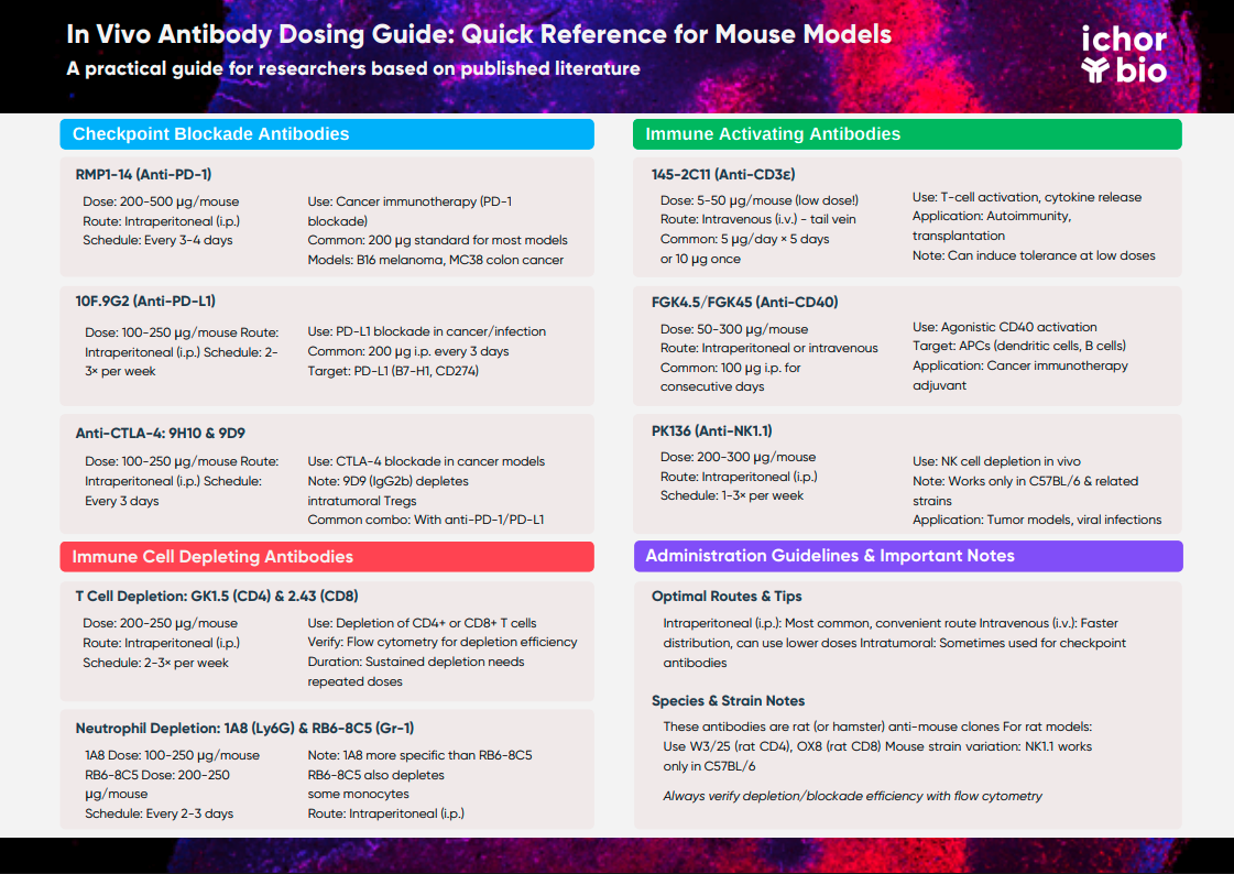
Find the Exact Antibody Dose You Need for Your Research
This comprehensive resource provides researchers with precise dosing information for commonly used antibody clones in mouse models. Whether you're conducting cancer immunotherapy studies with checkpoint inhibitors, depleting specific immune cell populations, or activating immune responses, this guide covers standard doses, administration routes, and experimental applications based on peer-reviewed literature.
Checkpoint Blockade Antibodies
How Much RMP1-14 (Anti-PD-1) To Use In Vivo
Target: PD-1 (CD279)
Species: Mouse
Standard Dose Range: 200-500 μg per mouse Recommended Route: Intraperitoneal injection
Optimal Dosing Schedule: Every 3-4 days
Applications: Cancer immunotherapy studies (PD-1 checkpoint blockade); reinvigorating exhausted T cells in chronic infection models
Published Examples: Often used at 200 μg per dose in syngeneic tumor models including MC38 and B16 melanoma
How Much 10F.9G2 (Anti-PD-L1) To Use In Vivo
Target: PD-L1 (B7-H1, CD274)
Species: Mouse
Standard Dose Range: 100-250 μg per mouse
Recommended Route: Intraperitoneal injection
Optimal Dosing Schedule: 2-3 times per week
Applications: Cancer immunotherapy studies (PD-L1 blockade); also used in infection models to enhance T-cell responses by blocking PD-1/PD-L1 interaction
How Much 29F.1A12 (Anti-PD-1) To Use In Vivo
Target: PD-1 (CD279)
Species: Mouse
Standard Dose Range: 100-200 μg per mouse (~5 mg/kg)
Recommended Route: Intraperitoneal injection
Optimal Dosing Schedule: Given 3 times at 3-day intervals
Applications: Cancer immunotherapy (PD-1 blockade); used in tumor models to stimulate anti-tumor T-cell activity, often in combination with other treatments
How Much 9H10 (Anti-CTLA-4) To Use In Vivo
Target: CTLA-4 (CD152)
Species: Mouse
Standard Dose Range: 100-200 μg per mouse
Recommended Route: Intraperitoneal injection
Optimal Dosing Schedule: Every ~3 days
Applications: Cancer immunotherapy (CTLA-4 blockade in tumor models to enhance T-cell activation; often combined with anti-PD-1/PD-L1 therapy; mimics ipilimumab's effect)
How Much 9D9 (Anti-CTLA-4) To Use In Vivo
Target: CTLA-4 (CD152)
Species: Mouse
Standard Dose Range: 100-250 μg per mouse
Recommended Route: Intraperitoneal injection (also tested intratumoral)
Optimal Dosing Schedule: Every 3 days
Applications: Cancer immunotherapy (CTLA-4 blockade); notably, clone 9D9 can deplete intratumoral Tregs due to its mouse IgG2b Fc, enhancing anti-tumor immunity
Immune Cell Depleting Antibodies
How Much GK1.5 (Anti-CD4) To Use In Vivo
Target: CD4 (T helper cell marker)
Species: Mouse
Standard Dose Range: 200-250 μg per mouse
Recommended Route: Intraperitoneal injection
Optimal Dosing Schedule: 2-3 times per week
Applications: CD4⁺ T cell depletion studies - used to test CD4 T cell requirement in autoimmune disease models, infection immunity, tumor rejection, and transplant tolerance
How Much 2.43 (Anti-CD8) To Use In Vivo
Target: CD8α (Cytotoxic T cell marker)
Species: Mouse
Standard Dose Range: 250 μg per mouse
Recommended Route: Intraperitoneal injection
Optimal Dosing Schedule: 2-3 times per week
Applications: CD8⁺ T cell depletion - eliminates cytotoxic T lymphocytes to assess their role in tumor immunity, viral clearance, graft rejection, etc. Often used alongside GK1.5 to deplete all T cell subsets
How Much RB6-8C5 (Anti-Gr-1) To Use In Vivo
Target: Gr-1 (Ly6G/Ly6C on neutrophils)
Species: Mouse
Standard Dose Range: 200-250 μg per mouse
Recommended Route: Intraperitoneal injection
Optimal Dosing Schedule: Every 2-3 days for sustained neutrophil depletion
Applications: Neutrophil/granulocyte depletion - used to study neutrophils' role in inflammation, infection, and cancer models
How Much 1A8 (Anti-Ly6G) To Use In Vivo
Target: Ly6G (neutrophil-specific marker)
Species: Mouse
Standard Dose Range: 100-250 μg per mouse
Recommended Route: Intraperitoneal injection
Optimal Dosing Schedule: 3 times per week or every 3 days
Applications: Neutrophil-specific depletion - more selective than RB6-8C5; used in tumor immunotherapy studies, infection models, and inflammation assays
How Much PK136 (Anti-NK1.1) To Use In Vivo
Target: NK1.1 (NK cell marker, also known as NKR-P1C/CD161)
Species: Mouse (NK1.1 is present in C57BL/6 and related strains)
Standard Dose Range: 200-300 μg per mouse
Recommended Route: Intraperitoneal injection
Optimal Dosing Schedule: 1-3 times per week as needed
Applications: NK cell depletion - eliminates natural killer cells to assess their function in tumor metastasis control, viral infections, and immune regulation
Immune Activating Antibodies
How Much 145-2C11 (Anti-CD3ε) To Use In Vivo
Target: CD3ε (T-cell receptor complex)
Species: Mouse
Standard Dose Range: 5-50 μg per mouse
Recommended Route: Intravenous injection (tail vein)
Optimal Dosing Schedule: 5 μg/day for 5 days or single 10 μg dose
Applications: Polyclonal T-cell activation in vivo, causing transient T-cell proliferation and cytokine release; used to induce T-cell depletion/activation-induced cell death and immune tolerance in autoimmunity
How Much FGK4.5/FGK45 (Anti-CD40) To Use In Vivo
Target: CD40 (costimulatory receptor on APCs)
Species: Mouse
Standard Dose Range: 50-300 μg per mouse
Recommended Route: Intraperitoneal or intravenous injection
Optimal Dosing Schedule: Single or consecutive daily doses
Applications: Agonistic CD40 activation to stimulate antigen-presenting cells; used as immune adjuvant in cancer immunotherapy, in vaccine models, and in autoimmune disease models
Administration Guidelines
Optimal Routes for In Vivo Antibody Administration
- Intraperitoneal (i.p.) injection: Most common route for these antibodies due to convenience and proven efficacy
- Intravenous (i.v.) injection: Used for faster or more uniform distribution (often allows slightly lower doses)
- Intratumoral injection: Has been explored in some cases for checkpoint antibodies to maximize local effect
- Dose timing: For sustained depletion or blockade, antibodies are typically administered every 2-4 days
- Initial dosing: Sometimes a higher "loading dose" is used, followed by lower maintenance doses
How to Verify Successful Antibody Treatment
Always confirm depletion/blockade efficiency in your specific model by flow cytometry or functional assays. Efficiency may vary by strain, antibody lot, or experimental conditions.
Species Specificity Notes
- The antibodies in this guide are primarily rat (or hamster) anti-mouse monoclonals used in mice
- For rat studies, different clones are typically needed as most of these don't cross-react with rat antigens
- Example rat-specific clones: W3/25 for rat CD4, OX8 for rat CD8, anti-rat NKR-P1 for NK cells
- Mouse strain variation: Some antibodies (particularly NK1.1) work only in specific mouse strains (e.g., C57BL/6)
- Clone 9D9 (anti-CTLA-4) is mouse IgG2b (not rat) and has special Treg-depleting properties in tumors
References
Checkpoint Blockade Antibody References
- AACR Journals: PARP Inhibitors Trigger the STING-Dependent Immune Response
- Journal for ImmunoTherapy of Cancer: The human IL-2 transgene promotes regulatory T cell expansion and protects mice from experimental autoimmune encephalomyelitis
- AACR Journals: Response to BRAF Inhibition in Melanoma Is Enhanced When Combined with Immune Checkpoint Blockade
- AACR Journals: Robust Antitumor Responses Result from Local Chemotherapy and CTLA-4 Blockade
- Journal for ImmunoTherapy of Cancer: Targeting interferon signaling and CTLA-4 enhance the therapeutic efficacy of anti-PD-1 immunotherapy in preclinical model of HPV+ oral cancer
- Cancer Biomedicine: Exploring the impact of therapeutic IgG1 and IgG2 anti-CTLA-4 on distinct anti-tumor responses and therapeutic potentials
Immune Cell Depleting Antibody References
- The Journal of Immunology: CD4+ T Cells Mediate IFN-γ-Independent Control of Mycobacterium tuberculosis Infection Both In Vitro and In Vivo
- The Journal of Immunology: Essential Role of Neutrophils in the Initiation and Progression of a Murine Model of Rheumatoid Arthritis
- The Journal of Immunology: NK Cells Alleviate Lung Inflammation by Negatively Regulating Group 2 Innate Lymphoid Cells
- Bioz.com: Anti-CD8 depleting antibody results
- JCI Insight: Neutrophil-mediated hypoxia drives pathogenic CD8+ T cell responses in cutaneous leishmaniasis
- Protocols.io: Natural killer cell depletion in vivo - mouse
Immune Activating Antibody References
- Clinical & Experimental Immunology: Inter-mouse strain differences in the in vivo anti-CD3 induced cytokine release
- Diabetes Journal: Oral Delivery of Glutamic Acid Decarboxylase (GAD)-65 and IL10 by Lactococcus lactis Reverses Diabetes in Recent-Onset NOD Mice
- AACR Journals: Combined Anti-CD40 and Anti–IL-23 Monoclonal Antibody Therapy Effectively Suppresses Tumor Growth and Metastases
- Blood Advances: Line-selective macrophage activation with an anti-CD40 antibody drives a monocyte-derived dendritic cell-like phenotype
Note: These references represent a selection of published studies that have used the antibodies at the doses and routes described in this guide. Individual experiments may require optimization based on specific models, mouse strains, and experimental goals.
Last updated: March 2025


